The gold standard for sleep monitoring and sleep disorders evaluation is PSG (Polysomnography). In this technique, the patient sleeps in a laboratory while his physiologic parameters are measured.
There is a rich diversity of PSG systems available, classified by AASM(American Academy of Sleep Medicine) in two levels: level 1 for the standard PSG and level 2 for portable PSG.
For a sleep study both levels must accommodate the minimum of seven channels:
- EEG(Electroencephalogram): to determine arousals from brain activity;
- EOG(Electroculogram): to detect REM sleep;
- EMG(Electromyography) submentonian: to look for limb movements that cause arousals;
- ECG(Electrocardiogram): for heart rate calculation
- ChestWall monitors: to document respiratory movements;
- Thermistor and/or nasal cannula: to nasal and oral airflow measurements;
- Oximetry: to measure oxyhemoglobin saturation(SpO2).
In terms of PSG home sleep testing, there are some solutions in the market. Somté PSG [1]implements a fully PSG home sleep testing. It has twenty five available channels and a software for data analysis.
Although offers a complete sleep study, such modality is not comfortable and is invasive for person in study as well as expensive, time-consuming and complex.
Recent studies have shown that heart rate alone has the potential to be a reliable mean for identifying sleep stages and diagnosing some sleep disorders. For example, it is a known fact that episodes of OSA are accompanied by a characteristic HR pattern consisting of bradycardia during apnea followed by abrupt tachycardia on its cessation [2], which can be used to detect OSA.
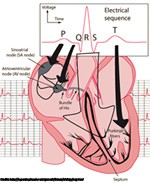
The electrocardiogram or ECG is a major diagnostic tool for the assessment of the health of the heart. It is a measurement taken at the surface of the skin which reflects the electrical phenomena in the heart when the SA (sinoatrial) node triggers the electrical sequence that controls heart action.
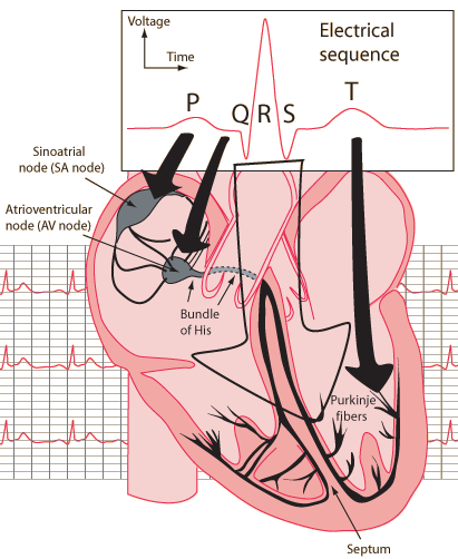
Figure 1- Schematic representation of normal ECG
Credits: http://hyperphysics.phy-astr.gsu.edu/hbase/biology/ecg.html
Normal heart beat consist of a P wave, a QRS complex and a T wave, Figure 1.
The P wave is the electrical signature of the current that causes atrial depolarization.
The QRS complex corresponds to the current that causes contraction of the left and right ventricles. This contraction is much more forceful than that of the atria and involves more muscle mass, thus the resulted ECG deflection is greater.
The T wave represents the re-polarization of the ventricles.
ECG signal is governed by autonomous nervous system. This is the reason why the ECG signal is well correlated with breathing [3] and can be source of the correlation with the different sleep stages [4] [5].
There are many methods for the processing of ECG signal. The four most documented methods are Heart Rate Variability (HRV) , Detrended Fluctuation Analysis (DFA) , Progressive Detrended Fluctuation Analysis (PDFA), Heart Rate Morphology ,Multi-scale Entropy Analysis, Information-Based Similarity.
There are some few difficulties in processing of the ECG signal which are imposed by its biological origin : Heart rate has many individual components and is driven by competitive forces (sympathetic and parasympathetic) and more over there are more of regulation mechanisms. This causes the creation of complex fluctuations. These fluctuations are not simply the result of responses on external factors, but they are persistent during physical load, rest and sleep. This non-stationarity is common for stochastic processes and therefore imposes that similar methods of processing may be used.
Recent studies have shown that HRV has the best results in the sleep disorders field.
HR(heart rate) can be derived directly from the ECG, or indirectly from other physiological waveforms, such as the PPG signal [6]. Analysis both in time domain and frequency domain are possible.
In a study from 2006 [7], HR variation was used to estimate REM and N-REM sleep durations, given that HR variation is well correlated with the sleep cycle [8] . A concordance of 88.8% was obtained for the estimated REM Sleep compared to actual REM sleep detected with EEG, EOG and EMG.
HRV(heart rate variability) is a black box with HR as the output. A study from 2011 [9], reviewed the ways in which HRV can be applied to understand autonomic changes during different sleep stages.It has also been applied to understand the effect of sleep-disordered breathing, periodic limb movements and insomnia both during sleep and during the daytime. HRV has been successfully used to screen people for possible referral to a Sleep Lab. It has also been used to monitor the effects of continuous positive airway pressure (CPAP).
A novel HRV measure, cardiopulmonary coupling (CPC) has been proposed for sleep quality. Evidence also suggests that HRV collected during a PSG can be used in risk stratification models, at least for older adults. Caveats for accurate interpretation of HRV are also presented. Conclusions of the study [9] clearly demonstrate the relevance of HRV analysis to clinical sleep medicine. At the same time, it states that clinical applications of HRV to sleep are still in their infancy.
Another study from 2008 [10] described an analysis algorithm of the ECG signal that is capable of diagnosing SBD by detecting episodes using CVHR(cyclic variations in heart rate). Applying the algorithm to 35 polysomnographic recordings provided by MIT Apnea Database the authors achieved a diagnostic accuracy of 77%.Using the ECG signal for detecting episodes of sleep disordered breathing seems reasonable as this events are known to be associated with autonomic reactions such as increases in blood pressure or frequent CVHR [11], but further improvements on the algorithm are needed.
The analysis of dynamic changes in ECG morphology using sophisticated algorithms enables the derivation of respiration from the ECG by tracking the amplitudes of the prominent R-wave. The derived respiratory curve is called ECG-derived respiration and correlates reasonably well with respiratory effort based on inductance plethysmography.
The combination of EDR(ECG-derived respiration) and sleep apnea related HRV has the potential to be a good and reliable detection tool for sleep apnea, but high-quality,empirical evidence on this subject is so far lacking [12].
References
|
[1] |
"COMPUMEDICS," 2014. [Online]. Available: http://www.compumedics.com.au/index.php/sleep-diagnostics/ambulatory/full-psg-ambulatory/somte-psg. |
|
[2] |
C. Guilleminault, R. Winkle, S. Connolly, K. Melvin and A. Tilkian, "Cyclical variation of the heart rate in sleep apnoea syndrome: mechanisms, and usefulness of 24 h electrocardiography as a screening technique," 1984. |
|
[3] |
S. R. Heinrich, F. Becker, S. Havlin, A. Bunde, J. W. Kantelhardt and T. Penzel, "Breathing during REM and NREM sleep: correlated versus uncorrelated behaviour," 2003. |
|
[4] |
J. W. Kantelhard, T. Penzel, J. Hermann, J.-H. Peter, A. Bunde, S. Havlin and K. Voigt, "Correlated and uncorrelated regions in heartrate fluctuations during sleep," 2000. |
|
[5] |
H. Becker, J. Peter, A. Bunde, T. Penzel and J. Kantelhard, "Detrended fluctuation analysis and spectral analysis of heart rate variability for sleep stage and sleep apnea identification," 2003. |
|
[6] |
J. Allen, "Photoplethysmography and its application in clinical physiological measurement," 2007. |
|
[7] |
M. Yaso, S. T. A. Nuruki and K. Yunokuchi, "Detection of REM sleep by heart rate," 2006. |
|
[8] |
M. Koki, M. Yaso, S. Tsujimura and K. Yunokuch, "Detection of the period of the REM sleep using HR in real time," 2005. |
|
[9] |
P. K. Stein and Y. Pu, "Heart rate variability, sleep and sleep disorders," 2011. |
|
[10] |
S. Canisius, T. Ploch, V. Gross, A. Jerrentrup, T. Penzel and K. Kesper, "Detection of Sleep Disordered Breathing by automated ECG analysis," 2008. |
|
[11] |
J. H. Peter, U. Koehler, L. Grote and T. Podszus, "Manifestations and consequences of obstructive sleep apnoea," 1995. |
|
[12] |
N. Collop, S. Tracy and V. Kapur, "Obstructive sleep apnea devices for out-of-center (OOC) testing: technology evaluation," 2008. |
